Currently, fluoroscopy, Computerized Tomography (CT), and ultrasound are used to support percutaneous needle interventions. However, they have some limitations.
Fluoroscopy shows live images of bones but does not show soft tissue structures. On the other hand, most CT scanners, despite presenting soft tissue structures perfectly, miss the live-imaging capabilities. CT-fluoroscopy is possible, but it generates a higher X-ray dose compared to normal fluoroscopy. In addition, needle path planning can only be done in the axial plane and the choice of trajectories is limited by the working area within the gantry tilt.
With CT, the patient must be moved in and out of the CT scanner (around 5-8 times, with at least 4 slices) to check the needle progression and maintain accuracy, and the gantry restricts access to the patient.
Ultrasound also has its limitations. Even if it is a good option for soft tissues and kidneys, as well as a non-invasive live-imaging solution, it can have limitations in large patients, especially when targeting deeper structures. The technique is an adjunct to the surgeon’s skills, experience, and technique, and sometimes even experienced users have trouble achieving good results repeatedly.
A miniature robot to improve targeting under live imaging
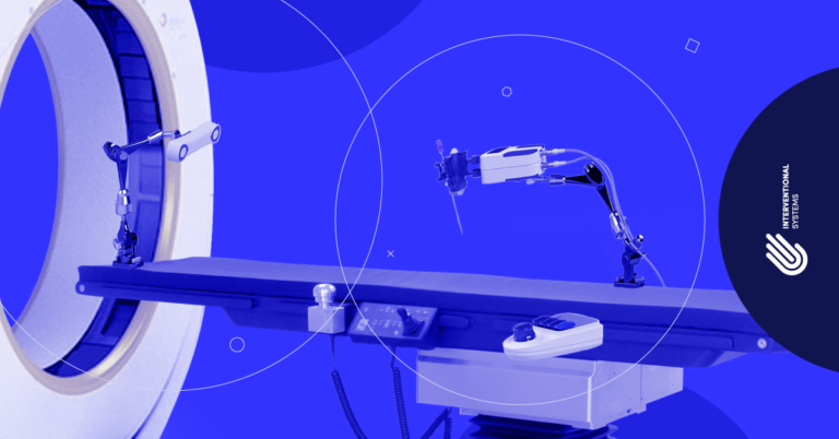
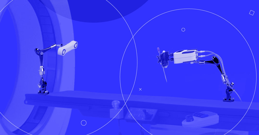
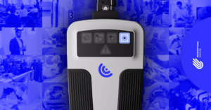
Micromate™ is a remotely controlled targeting device for image-guided interventions. The system is a robotic micro-positioning targeting platform that can be automatically or manually aligned to a surgical trajectory based on pre-operative or real-time 2D and 3D imaging. It is indicated for any percutaneous or minimally invasive procedure to which image-guided surgery is applicable.
The device is CE-Marked and obtained 510(k) clearance for usage in interventional radiology procedures under fluoroscopy, i.e., in Angio-CT and Cone-Beam CT (Angio/CBCT) suites. This combination is the only one that enables control under live X-ray imaging, real-time corrections if detected during instrument insertion, and submillimeter accuracy.
Even though hybrid rooms combining Angio/CBCT and CT devices are becoming ubiquitous, interventional radiology suites with a static or portable CT scanner remain the majority.
When performing a robotic-guided intervention using CT imaging, the robot would be mounted to the table, followed by a volume scan to achieve a registration, acquired with the patient and the robot in the range of interest. Based on this registration, the robot would then be capable to execute any planned trajectory within its range of motion.
Under certain circumstances, this approach can have a few flexibility drawbacks. If the robot needs to be re-positioned, the registration is no longer valid, and a new volume scan must be acquired. Moreover, the reachable anatomical volume is restricted by the robot’s range of motion. This increases radiation exposure, makes the workflow more cumbersome, and may limit intra-operative flexibility.
Combining Micromate™ with Atracsys spryTrack: more flexibility, same accuracy
At Interventional Systems, we believe that optical navigation can tackle the limitations encountered in typical CT-guided interventions. But creating a robotic navigation system is much more than combining a robot with a commercially available camera and displaying the data in a tracking software. That is the easy part.
A seamless integration in the clinical workflow, though, requires that we think about camera positioning, setup flexibility, occlusions, and ensuring the highest accuracy while almost even forgetting the camera is there. As with Micromate™, footprint and setup flexibility are key.
That’s why we have partnered with Atracsys. Their compact and versatile mobile optical tracking system, spryTrack 180, offers high accuracy and precision, while enabling innovative developments and making the integration smooth and seamless.
The spryTrack is mounted on the table, facing Micromate™ and the surgical site directly. This approach not only ensures the highest accuracy and allows for an intra-operative automatic registration, but it also removes the known field-of-view problems related to occlusions from the surgical staff and other bulky equipment, particularly when cameras are positioned in the ceiling or on its own cart.
More than just being combined, Interventional Systems and Atracsys’s technologies are integrated.
If you want to learn more about how this integration can help you in your applications, contact us here.
