Blog
How Micromate™ Made a Difference: The Story of Carlos Tavares
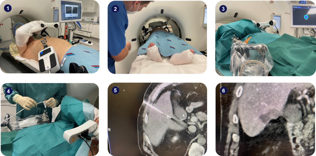
Carlos Tavares, 76, was one of the first patients to benefit from the robotic precision of Micromate™ in Portugal, at the Local Health Unit of Gaia and Espinho (ULS Gaia and Espinho).